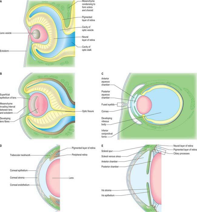
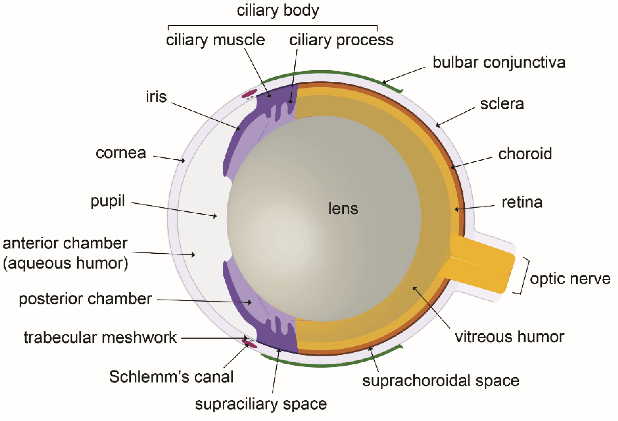
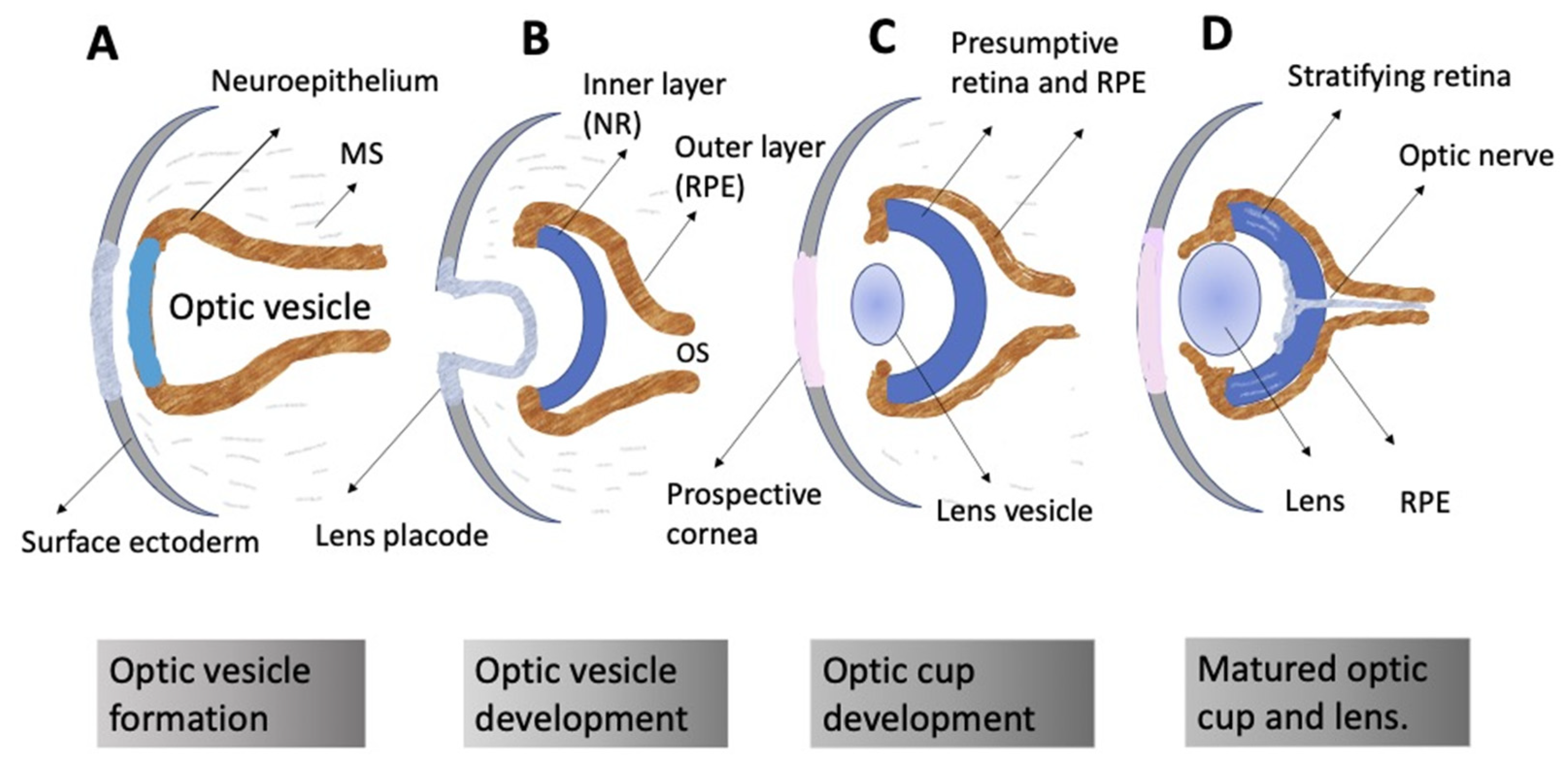
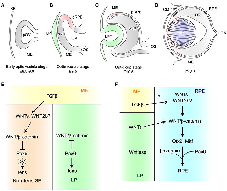
Schematic of eye development in frog, mouse and human; the eye develops is one of the most viral images on the internet at the moment. As well as to those photographs, Mouse eye diagram. Cross-sectional schematic of the human and mouse eye, Schematic Diagram Of Mouse Eye Development – Circuit Diagram and Mousetap technique. (a) Diagram of mouse eye showing aqueous humour also experienced a major increase in search traits. In case you are in search of trending photos related to Schematic of eye development in frog, mouse and human; the eye develops, on this web page we have now 35 images so that you can explore. Here you go:
Schematic Of Eye Development In Frog, Mouse And Human; The Eye Develops

www.researchgate.net
frog cells neural plate develops embryo optic
Schematic Diagram Of Mouse Eye Development – Circuit Diagram

www.circuitdiagram.co
Development Of The Eye | Basicmedical Key

basicmedicalkey.com
vessels diaphragm appendages basicmedicalkey
Schematic View Of Human And Mouse Eye (A) And Cross-section Of The

www.researchgate.net
The Use Of Confocal Laser Microscopy To Analyze Mouse Retinal Blood

www.intechopen.com
mouse confocal microscopy vessels retinal blood laser analyze use intechopen figure
Mousetap Technique. (a) Diagram Of Mouse Eye Showing Aqueous Humour

www.researchgate.net
aqueous technique lens vitreous
A Schematic Diagram Of Early Mouse Embryogenesis Showing The Lethal

www.researchgate.net
embryogenesis lethal ectoderm extraembryonic embryonic endoderm exe icm epc visceral trophectoderm complementation eme
Stages Of Lens Formation In Mouse Embryos. Schematics Showing The

www.researchgate.net
lens schematics embryos
Figure 1 From Cell Fate Decisions And Axis Determination In The Early

www.semanticscholar.org
Biology | Free Full-Text | Lymphatics In Eye Fluid Homeostasis: Minor

www.mdpi.com
Biomedicines | Free Full-Text | Self-Organization Of The Retina During

www.mdpi.com
Map Of Mouse Iris Offers New Look At The Eye | HHMI

www.hhmi.org
Pupil Dilation Deficits In Aged Gba KI Mice Are Apparent Post Mortem

www.researchgate.net
Comparative Overview Of Human And Drosophila Eye Development. Eye

www.researchgate.net
drosophila human development comparative embryonic imaginal genetics patterning
The Mouse Eye And Retina. A) The Mouse Eye Is Similar In Structure To

www.researchgate.net
retina similar retinal
Eyelid Development Of The Mouse At Embryonic Day 11.5 (E11.5) (A-A

www.researchgate.net
Eye Development In Vertebrates | Download Scientific Diagram

www.researchgate.net
(a) Schematic Diagram Of Murine Eye Development From Embryonic Days

www.researchgate.net
development embryonic murine retinal embryo e11 neural e10 tissue
Mouse Models Of Childhood Cancer Of The Nervous System | Journal Of

jcp.bmj.com
retinoblastoma journal nervous jcp bmj
Persona A Cargo Del Juego Deportivo Preámbulo Sexual Retina Mouse Cerca

mappingmemories.ca
Schematic Diagram Depicting The Developmental Regression Of Hyaloid

www.researchgate.net
Frontiers | WNT/β-Catenin Signaling In Vertebrate Eye Development

www.frontiersin.org
eye development wnt vertebrate signaling catenin optic vesicle frontiersin figure fcell
Mouse Eye Diagram. Cross-sectional Schematic Of The Human And Mouse Eye

www.researchgate.net
sectional figure source
(PDF) Stem Cell Function In The Mouse Corneal Epithelium

www.researchgate.net
embryonic
Scientists Use Gene Therapy And A Novel Light-sensing Protein To

www.nei.nih.gov
retina opsin mice vision location cell bipolar light therapy eye gene structure protein sensing restore novel scientists use layers diagram
Morphogenesis Of The Anterior Segment In The Zebrafish Eye. Comparison

www.researchgate.net
zebrafish embryonic segment morphogenesis factors simplified genetic forming histology dpf soules
Lens Placodes : Pax6 Activity In The Lens Primordium Is Required For

ayoobelajarcss.blogspot.com
vertebrate placodes primordium optic placode ucdavis brain ophthalmology
Mouse Eye Diagram. Cross-sectional Schematic Of The Human And Mouse Eye

www.researchgate.net
vitreous retina sectional lens jove マウス evisceration relative proteomic analyses 2795 protocol 断面 separate
Cross-sectional Diagram Of The Mouse Eye Highlighting Features

www.researchgate.net
sectional highlighting protocol referenced
Schematic View Of Human And Mouse Eye (A) And Cross-section Of The

www.researchgate.net
Retina similar retinal. Figure 1 from cell fate decisions and axis determination in the early. Vitreous retina sectional lens jove マウス evisceration relative proteomic analyses 2795 protocol 断面 separate